This article reveals the Biology of Sponges (phylum Porifera) by studying the Morphology, Anatomy, Histology, and Physiology of Sponges. The author has also included the Reproduction and Development of Sponges.
Porifera is a phylum comprising of the multi-cellular invertebrate animals called Sponges. The term “Porifera” literally means “Pore Bearers”. The animals of this phylum have tiny pores in their body walls, and this characteristic feature is the basis of the name of this phylum.
Porifera includes very primitive multi-cellular animals having only the cellular level of body organization with no tissues and organs. In Porifera (sponges), only cells show division of labor for the purpose of performing specialized functions. All Poriferans, animals of the phylum Porifera, are aquatic with most of them being marine. Sponges are sessile (not mobile) organisms including both solitary and colony-forming types.
Morphology and Canal System
Body of a sponge is vase-like, tubular (tube-like), cylindrical, branched, cushion-shaped, etc. Sponges are either radially symmetrical or have no symmetry (asymmetrical). The surface of the body bears pores known as Dermal Pores or Ostia (singular is “Ostium”, which in Latin means: door). The pores lead directly or indirectly into the central cavity known as Spongocoel (spongos, sponge + koilos, cavity) or Paragastric Cavity. This cavity opens to the outside through a pore (or pores) known as Osculum (or Oscula).
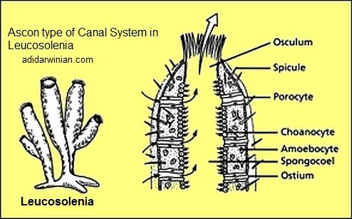
Ascon type of Canal System
In some sponges, like Leucosolenia, just these three components: Ostia, Spongocoel, and Osculum – form the canal system. This is the simplest type and is called the Ascon type of canal system. In this type of canal system, water enters from outside through Ostia into the Spongocoel, and then, leaves through the Osculum to the exterior. Sponges having Ascon type of canal system are known as Asconoid Sponges.
 Flow of water in Ascon type of canal system:
Water flows from outside (through Ostia) to Spongocoel (through Osculum) to outside.
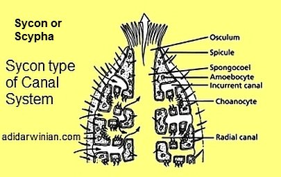
Sycon type of Canal System
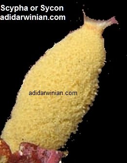
In other sponges, like Scypha (Sycon or Urn Sponge or Crown Sponge), folding of the body wall into finger-like processes occurs. Body wall folds to form alternating invaginations (Incurrent Canals) and evaginations (Radial Canals). This type of complex system, comprising of Ostia, Incurrent Canals, Radial Canals, Spongocoel, and Osculum, is called Sycon type of canal system. Sponges having Sycon type of canal system are called as Syconoid sponges, for example, Scypha (Sycon), Grantia, etc.

Flow of water in Sycon type of canal system:
Water flows from outside (through Ostia) to Incurrent Canal (through spaces called Prosopyles) to Radial Canals (through openings called Apopyles) to Spongocoel (through Osculum) to outside.
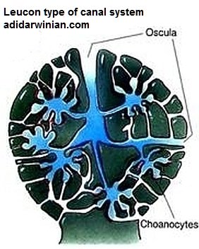
Leucon type of Canal System
Further folding of the body wall leads to the formation of a more complex type of canal system known as the Leucon type of canal system. In this type, Incurrent Canals open into Flagellated Chambers through the Prosopyles. There is no Spongocoel, and instead the Excurrent Canals are present. The Flagellated Chambers open into the Excurrent Canals through the Apopyles. The Excurrent Canals open to the outside via the Osculum. Sponges having Leucon type of canal system are called as Leuconoid sponges, for example, Plakina, Geodia, Spongilla, Oscarella, etc.
 Flow of water in Leucon type of canal system:
Water flows from outside (through Ostia) to Incurrent Canals (through spaces called Prosopyles) to Flagellated Chambers (through openings called Apopyles) to Excurrent Canals (through Oscula) to outside.
Body Wall
The body wall, which encloses the Spongocoel, consists of three layers, viz. –
Pinacoderm – The external surface of the body of Leucosolenia, except the Osculum and Dermal Pores, is covered by a cellular layer called Pinacoderm, which is composed of Pinacocytes (pinaco, plank+ kytos, cell). In Scypha (Sycon), the layer of Pinacocytes covering the outer body surface, except the Osculum and Dermal Pores, is called Exopinacoderm. The layer of Pinacocytes lining the incurrent canals and Spongocoel in Sycon is called Endopinacoderm.
Mesenchyme – The layer lying between the Pinacoderm and Choanoderm is non-cellular and is called the Mesenchyme. It is made up of a gelatinous matrix and contains several types of amoeba-like cells called Amoebocytes (Amebocytes). Amoebocytes include the following types of cells: Archaeocytes, which can produce all other types of Amoebocytes; Chromocytes (pigmented amoebocytes); Collencytes (have branching pseudopodia forming a network); Thesocytes (have rounded or lobose pseudopodia and food reserve); Scleroblasts (spicule-forming amoebocytes); Myocytes (muscle cells surrounding Osculum and Ostia); Gland Cells (secrete lime); and Germ Cells (sperms and ova).
Choanoderm – Choanoderm consists of flagellated collar cells known as Choanocytes (choane, funnel + kytos, cell). This cellular layer lines the Spongocoel in the most primitive type of sponges (like Leucosolenia). The radial canals and flagellated chambers are lined by Choanoderm. Choanoderm is also called Gastrodermis or Gastral Epithelium. Choanocytes are involved in feeding, and through beating of their flagella ensure the flow of water within the body of a sponge.
Sponges are not truly diploblastic because cells are loosely arranged and do not form definite layers.
Skeleton of Sponges
Skeleton of Sponges (Poriferans) include Spicules, or Spongin fibers, or both. Skeleton is embedded in the Mesenchyme. It protects and supports the soft parts of a sponge’s body. Skeleton helps in the classification of Sponges.
Spicules
Spicules (spica in Latin means point) are crystalline structures having rays or spines. They are secreted by Scleroblasts of Mesenchyme. Spicules are calcareous (consisting of Calcium Carbonate or Calcite; these spicules occur in the class Calcarea of phylum Porifera) or siliceous (consisting of colloidal Silica or Silicon; these spicules occur in the class Hexactinellida of phylum Porifera) in nature. Spicules, embedded in the Mesenchyme, form the endoskeleton in order to protect and support the soft body parts. Monaxon spicules are straight or curved and needle-like or rod-like structures that grow along a single axis. Tetraxon spicules consist of four rays in different planes. Spicules having three axes crossing one another at right angles to form six rays are termed as Triaxon spicules. Polyaxon spicules have several equal rays radiating from a central point. Spicules projected from the body surface form bristles. Spicules surrounding the Osculum form the Oscular Fringe.
Spongin FibeRs
Spongin fibers occur in class Demospongiae of phylum Porifera. Spongin fibers consist of a Sulphur containing scleroprotein called Spongin. Spongin also contains Iodine, the amount of which is larger in certain tropical species of Spongiidae and Aplysinidae. It is worth noting here that the herbal doctors, in the old times, used Bath-Sponge (Spongia / Euspongia belonging to the family Spongiidae) for the treatment of Croup (click here to see what is Croup?).
Respiration and Nervous System
There are no special respiratory organs, and respiration takes place by the diffusion of gases between the cells and flowing water. In Porifera, nervous system is absent or primitive.
Nutrition and Digestion
Digestion is intracellular. Microscopic organisms (planktons) and organic particles enter the body with water through the Dermal Pores or Ostia. Food is taken up by the food vacuoles of Choanocytes. Partly digested food is then passed to the Amoebocytes, where the digestion is completed. Amoebocytes distribute the digested food to other cells.
Excretion
There are no special excretory organs, and excretion of metabolic wastes (chiefly Ammonia) takes place by diffusion into the outgoing water current.
Reproduction
Both asexual and sexual methods of reproduction are found in phylum Porifera. Sponges exhibit high power of regeneration.
Asexual Reproduction
Asexual reproduction occurs by the following methods:
Budding: In Budding, an evagination of the body or outgrowth from the body occurs near the base of the body in order to form a bud. Fully grown bud may remain attached with the parent as a part of the colony or gets detached to form a new sponge.
Branching: In Branching, new horizontal branches arise from the older horizontal tubes or stolons and give rise to erect branches. An erect branch or upright cylinder breaks-off an Osculum at its free end, and thus, becomes the part of the colony.
Fission: A limited area of sponge gets hypertrophied and develops a line of weakness. Fission or splitting occurs along the weak line, thus, separating a part from the parent body. This part develops into a new individual.
Reduction Bodies: In this unusual method of asexual reproduction, sponge disintegrates during the adverse conditions into small rounded balls known as the reduction bodies. Each reduction body consists of a mass of Amoebocytes covered by Pinacoderm. On the return of favorable conditions, reduction bodies grow into new sponges.
Gemmules: Internal buds or gemmules (gemma means a bud) are formed by all freshwater and some marine sponges as a method of asexual reproduction. In autumn, sponges suffering from the unfavourable conditions, like cold or scarcity of food, disintegrate into a large number of gemmules. Gemmules can withstand freezing conditions of winter, and under favourable conditions of spring, develop into new sponges. New sponges give rise, through sexual reproduction, to summer generation of sponges that again die in autumn leaving the gemmules. Thus, the life history of these sponges exhibits alteration of generations.
Sexual Reproduction
Sponges are hermaphrodite (click here to see what is Hermaphrodite?) but still the reproductive process involved is cross-fertilization only. There are no special sex organs. Sperms (male reproductive gametes) and ova (female reproductive gametes) are formed in the same individual from archaeocytes, or from choanocytes.
Spermatogenesis: A sperm mother cell (Spermatogonium), which is an enlarged archaeocyte or a modified choanocyte, gives rise to Spermatocytes that form Spermatozoa (male sex cells or sperms). The process of the formation of haploid gametes from diploid cells by meiosis is termed Gametogenesis (Spermatogenesis – if sperms are formed; Oogenesis – if ova are formed). Spermatozoa or sperms are released from the Mesenchyme into the sea water.
Oogenesis: An egg mother cell (Oocyte), which is an enlarged archaeocyte or a modified choanocyte, gives rise to Ovum (female sex cell). Ova remain in the Mesenchyme.
Fertilization: The sperms released into the sea water by an individual enter into other sponges and fertilize the ova present in the Mesenchyme of the body wall (in situ fertilization). To know about the term in situ, see what is in situ? Thus, cross-fertilization occurs and the process of fertilization is internal (internal fertilization). The fusion of a sperm and an ovum leads to the formation of the Zygote.
Development
Early development of the zygote takes place in maternal sponge resulting in the formation of a larva. With the help of flagella, the larva escapes from the maternal body into the outside water. After swimming, for some hours to many days, it settles down, attaches to a solid object, and undergoes metamorphosis (transformation of a larva into an adult). Two types of larvae occur in sponges:
Amphiblastula
It occurs in many calcareous sponges, for example, Scypha (Sycon). It is hollow, oval, and only its anterior one-half part bears flagella.
Parenchymula
It occurs in some sponges of class Calcarea, Hexacctinellida, and most Desmospongiae. It is solid, oval, or flattened, and its entire outer surface bears flagella.


Sponges are one of the excellent examples of Mother Nature’s marine life creation.
I totally agree.
This is excellent information on sponges.
Pingback: Katy
Well-researched and beautifully illustrated article on Sponges!!
Yeah, I too agree with you Loney.
Great work, the biology of beautiful animals called sponges revealed in a readily intelligible manner!