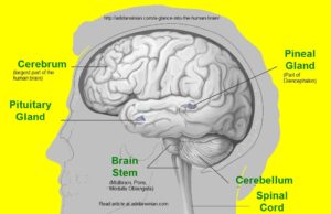
A Glance into the Human Brain – Structure and Functions of the Human Brain (Human Brain’s Anatomy and Physiology) – A Voyage Into the Human Brain!
This article describes the structure of human brain (anatomy of human brain) and functions of human brain (physiology of human brain), which is the most complex and mysterious organ of the human body. This article is expected to cater the needs of a variety of audiences including the students, teachers, healthcare professionals, and the laymen. The article has been written in an easy-to-understand language and richly annotated with definitions of difficult medical terms including etymologies.
The human brain is the most sophisticated organ of the human body, and can aptly be called as a biological marvel. The brain and the spinal cord constitute the central nervous system (CNS). The brain plays the role of the control tower or control center of the human body, and relies on a vast network of nerves (bundles of fibers of nervous tissue carrying impulses) spread throughout the body. Nerves can be compared to the electrical wiring as they carry electrical impulses.
Noteworthy Facts about the Human Brain!
- The brain of human beings resembles a small cauliflower in its size and appearance.
- Human brain is comprised of about 100 billion neurons or nerve cells.
- The weight of an adult human brain is about 1300 g (about 3 pounds), whereas the weight of a newborn’s brain lies in the range of 350 – 400 g.
- Although the brain of a human being accounts for only 2 % of the total body weight, it utilizes 20 % of the of the resting total body oxygen consumption.
- Human brain contains 77 to 78 % water, 10 to 12 % lipids, 8 % proteins, 2 % soluble organic substances, 1 % carbohydrates, and 1 % inorganic salts.
- The cerebral cortex forms 77% by volume of the human brain.
- Cerebrum is the largest part of the human brain, whereas Cerebellum is the second largest.
- Left side of the brain (left cerebral hemisphere) controls the right side of the body, whereas the right side of the brain (right cerebral hemisphere) controls the left side of the human body.
Neurons in the Human Brain
There are about one-hundred billion nerve cells or neurons in the human brain. The nerve cells or neurons receive and transmit information with incredible speed and precision. A neuron consists of dendrites (branching processes) that receive signals or electrical impulses and transfer them towards the neuronal cell body (nerve cell body), a cell body, and an axon (a long and single fiber) that transmits information or signals away from the cell body to another neuron. The neurons interconnect with other neurons, thus forming the neural circuits. Neural circuits perform specific functions. The human brain possesses the ability to modify the connections between the neurons to adapt to new circumstances, learn, and memorize. This process is known as the brain plasticity.
The junction where a signal or impulse passes from one neuron to another neuron or cell is known as the synapse. To know more about synapse, see – what is synapse? The signal travels through the axon and reaches the end of it. At the end of the axon, the signal stimulates the release of tiny sacs containing chemicals. These chemicals, which are released into the synapse, are called the neurotransmitters. The molecules of a neurotransmitter cross the synapse and bind to the specific cell receptors present on the adjacent cell. The receiving cell then responds to the signal. If the receiving cell is a nerve cell, then this cell may again transmit the signal to the next cell. This way transmission of information occurs between the neurons in the brain.
Parts of the Human Brain
The human brain consists of four main parts, viz. – cerebrum, diencephalon, cerebellum, and brainstem.
Cerebrum
Cerebrum (Latin – brain) is the largest part of the human brain. The cerebrum is the topmost part of the human brain and is the source of a person’s intellectual activities. Cerebrum is aptly called the seat of intelligence. It enables one to think, reason, remember, learn, judge, will, imagine, plan, read, write, speak, calculate, and create. The sensory areas of the cerebrum receive and interpret impulses; the motor areas control muscular movements; and the association areas deals with the emotions, personality traits, and the intellectual processes. The cerebrum comprises of two cerebral hemispheres, each controlling one side of the human body. The inner core of cerebral hemispheres is made up of the white matter (myelinated nerve fibers; the white color is due to myelin) and the outer layer is made up of the gray matter (composed of cell bodies of neurons and unmyelinated fibers). This outer layer is called the cerebral cortex. Most of the actual processing of information in the human brain occurs in the cerebral cortex. Corpus callosum [hard body – body (corpus) + hard / tough (callosum)], a broad band of axons, connects the two cerebral hemispheres.
Cerebrum also contains the basal ganglia (ganglia is pleural of ganglion; in Greek, “ganglion” means a tumor or knot). The basal ganglia are comprised of several pairs of nuclei. In neurology, a single cluster or group of cell bodies of neurons (neuronal cell bodies) within the Central Nervous System (CNS) is known as a nucleus (plural – nuclei). Nuclei are devoid of myelin, and are thus, gray in color. The two members of each pair of the basal ganglia are oppositely located in the cerebral hemispheres of the cerebrum. Corpus striatum [striated body / striped body – body (corpus) + striated / striped (striatum)] is the largest nucleus in the basal ganglia. Basal ganglia are involved in the regulation of muscle tone and coordination of the automatic movements of skeletal muscles such as laughing in response to something funny.
Diencephalon
Diencephalon (literally means “through brain”) lies below the cerebrum and above the brain stem. It consists of thalamus, hypothalamus, epithalamus, and subthalamus.
Thalamus (from Greek “thalamus”, which means “inner chamber”) acts as the principal relay station for sensory impulses reaching the cerebral cortex.
Hypothalamus lies below the thalamus. Hypothalamus produces many hormones including oxytocin and vasopressin (antidiuretic hormone). Hypothalamus also controls the pituitary gland (also known as hypophysis). Hypothalamus is one of the principal regulators of homeostasis in human body. Hypothalamus controls autonomic nervous system; body temperature; thirst; appetite and satiety; feelings of pleasure, fear, rage, and pain; sexual behaviors including sexual arousal, mating, and child rearing; and sleeping and waking cycles.
Epithalamus lies above the thalamus. It consists of the pineal gland (secretes melatonin) and clusters of neuronal cell bodies (nuclei).
Subthalamus lies immediately below the thalamus and is involved in the control of body movements.
Cerebellum
Cerebellum (Latin – a small brain) is the second largest part of the human brain. Cerebellum is present under the cerebrum at the back of the head. Cerebellum looks like a butterfly in shape. It consists of two cerebellar hemispheres (representing the wings of a butterfly) separated by a central narrow strip-like area known as the vermis (in Latin, “vermis” means “worm”). Cerebellum controls and coordinates voluntary muscle movements by sending feedback to the motor areas of cerebrum. Cerebellum helps to make movements of the voluntary muscles smooth and precise. Cerebellum regulates posture and balance of the body. Cerebellum also controls skilled muscular activities like dancing.
Brain Stem
The area of the brain lying between the spinal cord and the diencephalon constitutes the brain stem. The brain stem is comprised of midbrain, pons, and medulla oblongata.
Midbrain
Midbrain, also known as the mesencephalon, is the uppermost part of the brainstem. It consists of groups of cell bodies (nuclei) and nerve fibers (tracts). Several nuclei are present in the midbrain including the substantia nigra [substantia (substance) + nigra (black)] and the red nuclei. Substantia nigra consists of large darkly pigmented nuclei controlling subconscious muscle activities. The red nuclei have rich blood supply and iron-containing pigment. The red nuclei are involved in the coordination of muscular movements. Nuclei in the midbrain control the movements of the eyeball via the following cranial nerves, viz. – oculomotor nerves and trochlear nerves. Midbrain also has reflex centers that control movements of the eyes, neck, head, and trunk in response to stimuli. The anterior part of the midbrain consists of a pair of tracts known as cerebral peduncles. These tracts contain axons of motor and sensory neurons.
Pons
Pons (Latin, bridge) is present below the midbrain, in front of the cerebellum, and above the medulla oblongata. Pons is also known as the bridge of Varolius or pons Varolii after the anatomist Costanzo Varolio (Constantius Varolius). The transverse nerve fibers (tracts) of pons form a bridge between the right and left cerebellar hemispheres, and the longitudinal fibers are part of the ascending and descending tracts between the brain and the spinal cord. Nuclei present in the pons help control breathing. Pons also has some nuclei that are associated with the following four pairs of cranial nerves, viz. – trigeminal nerves, abducens nerves, facial nerves, and vestibulocochlear nerves.
Medulla Oblongata
Medulla Oblongata, or Medulla, forms the lowermost part of the brainstem. It extends from the pons above to the spinal cord below. Medulla has many nuclei that regulate various vital functions. Medulla also has nuclei that are associated with the following five pairs of cranial nerves, viz. – vestibulocochlear nerves, glossopharyngeal nerves, vagus nerves, accessory nerves, and hypoglossal nerves. Medulla also has nuclei involved in the control of various autonomic functions. These nuclei include the medullary rhythmicity area of the respiratory center that regulates the basic breathing rhythm; the cardiovascular center (cardiac center) that controls the rate and force of the heartbeat; the vasomotor center that controls the diameter of the blood vessels; and the reflex centers that initiate the reflex actions of vomiting, coughing, and sneezing.
The ascending tracts (sensory tracts) and the descending tracts (motor tracts) pass through the medulla oblongata. These tracts connect the spinal cord with the brain. The largest motor tracts passing from the cerebrum to the spinal cord form two pyramids in the medulla oblongata. Just above the junction of the medulla oblongata with the spinal cord, the crossing of pyramids or decussation of pyramids occurs, where most of the axons in the left pyramid cross over to the right side and most of the axons in the right pyramid cross to the left side. Due to this crossing of pyramids, the left cerebral cortex controls the skeletal muscles on the body’s right side and the right cerebral cortex controls the skeletal muscles on the body’s left side.
The sensory tracts (ascending tracts), connecting the spinal cord with the brain, decussate (cross over) in the spinal cord (anterior spinothalamic tract and lateral spinothalamic tract decussate in the spinal cord), or in the medulla oblongata (dorsal column-medial lemniscus pathway (DCML) / Posterior column-medial lemniscus pathway (PCML) decussates in the medulla oblongata). Therefore, the sensory information from the body’s left side is received by the right side of the brain and the sensory information from the body’s right side is received by the left side of the brain. Other sensory tracts (spinocerebellar tracts) transmit information from the muscles to the cerebellum directly without any crossing over.
Other Parts of the Human Brain
Reticular Formation
A net like region of interspersed grey and white matter is also present in the brain stem and is known as the reticular formation. Reticular (derived from Latin “reticulum”, which means “little net”) formation has both sensory and motor functions. It alerts the cerebral cortex to incoming sensory signals and also regulates muscle tone.
Limbic System
On the inner border of the cerebrum and floor of the diencephalon, are present a ring of structures known as the limbic system. Limbic (“Limbic” means border) system is also known as the emotional brain as it plays a principal role in a range of emotions. Limbic system consists of hypothalamus, amygdala, hippocampus, and other structures. The word hippocampus is derived from the Greek “hippokampos”, hippos (horse) + kampos (a sea monster); hippocampus is the Latin name of seahorse (a type of fish). In the human brain, hippocampus is a curved, elongated, elevated and seahorse-resembling structure of the limbic system, which is composed of gray matter covered by a layer of white matter. Amygdala, also known as amygdaloid nucleus or amygdaloid body, is an almond-shaped mass of gray matter (nucleus). A pair of amygdalae occurs in the human brain as a part of the basal ganglia.


Excellent synopsis of brain structure and function
Excellent brain article for the students of life sciences.
In-depth article; I especially liked the way of describing brain structure and functions along with the definitions of difficult medical terms.